Pericardial Effusion: Diagnosis And Treatment
The main means of diagnosing a pericardial effusion is the echocardiogram. We tell you everything about this disease below.
Pericardial effusion is a problem that is found relatively frequently in medical practice. Sometimes it is related to a known disease and other times it requires specific evaluation and follow-up to establish its cause.
Sometimes it is impossible to determine the cause of the pericardial effusion. This condition can remain unchanged for years, and become chronic without producing any hemodynamic compromise.
Initially, treatment focuses on solving the problem that gives rise to the pericardial effusion and managing its symptoms. However, when the cause is unknown, the clinical management that is given is the same as that of pericarditis.
What is pericardial effusion?
Pericardial effusion is defined as an abnormal accumulation of fluid in the pericardial cavity. Remember that the pericardium is made up of two layers: a visceral (internal) and a parietal (external).
The space between these two layers is the pericardial cavity or pericardial sac. Under normal conditions, it contains up to 50 milliliters of serous fluid. When there is an inflammatory or infectious process, fluid production increases and pericardial effusion occurs.
Likewise, this can be caused by the decrease in reabsorption of the liquid. This occurs due to the increase in systemic venous pressure. In turn, the increase in pressure generally occurs due to congestive heart failure or pulmonary hypertension.
Diagnosis
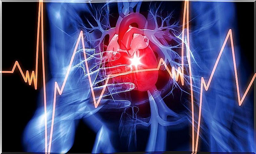
The clinical presentation of pericardial effusion depends on the speed with which the fluid accumulates. Typical symptoms are shortness of breath and chest pain. Nausea, dysphagia, hoarseness, and hiccups are also common.
When pericardial effusion is suspected, one or more of these tests are usually performed:
- Echocardiography. It allows to detect the magnitude of the effusion and the state of cardiac function. The transesophageal echocardiogram offers more detailed, and therefore more reliable, images than the subthoracic echocardiogram.
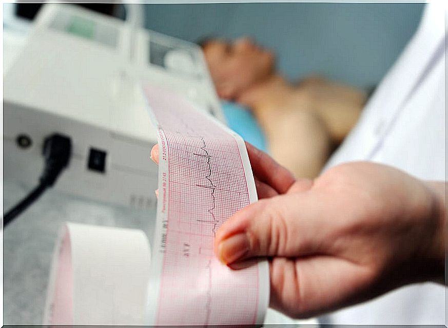
- Electrocardiogram. This test can detect patterns of possible plugging.
- Chest X-ray. It offers an indicator to establish the magnitude of the pericardial effusion globally.
Although the most widely used diagnostic test is the echocardiogram, computed tomography (CT) and magnetic resonance imaging (CMR) offer a wider field of vision. However, due to availability and costs, these tests are only used rarely.
In any case, the echocardiographic evaluation allows determining five fundamental variables: size, time of evolution, distribution, composition and hemodynamic effects. The next clinical challenge is to establish the cause of the pericardial effusion to proceed to define the line of treatment.
Treatment for pericardial effusion

The treatment of pericardial effusion depends directly on the amount of accumulated fluid, the existence or not of cardiac tamponade and the cause that gives rise to this anomaly. In general, treating the cause fixes the problem.
The first step in the management of a pericardial effusion is to assess its size, define its hemodynamic significance, and establish possibly associated diseases. In about 60% of cases, there is an underlying disease.
If there is no tamponade or a significant risk of it occurring, bed rest and anti-inflammatory drugs are usually ordered . It is also used withchicine and corticosteroids.
If there is tamponade or a high risk that the effusion progresses, it is indicated to carry out a pericardiocentesis. When it is not possible to perform it, or this failure, it is appropriate to carry out an open surgical drain. This should include biopsy and the creation of a pericardial window.
Monitoring and prognosis
In general, idiopathic pericardial effusion and pericarditis have a good prognosis. The risk of complications is very low. Chronic idiopathic effusions have a 30% to 35% chance of escalating to cardiac tamponade.
In the other types of effusion, the prognosis depends mainly on the cause that produces it and the size of the effusion. Those that are greater than 10 mm worsen and progress to tamponade in up to a third of all cases.
In moderate idiopathic effusions, it is advisable to do a follow-up that includes an echocardiogram every six months. If these are severe, it should be done every three months. And in the case of non-idiopathic patients, the follow-up will depend on the disease that gives rise to this anomaly.